
For laboratory research only. Not for human or animal use. This article gives no dosing, administration or therapeutic advice.
Short answer: endotoxin is lipopolysaccharide (LPS) from the outer membrane of Gram-negative bacteria. Cells sense it through the receptor TLR4[1], so even trace contamination can trigger inflammatory signalling and be mistaken for an effect of the peptide being studied[2][3]. The standard tests are Limulus amebocyte lysate (LAL) assays and recombinant Factor C (rFC)[9][12]. A routine peptide COA (HPLC purity plus mass-spectrometry identity) does not measure endotoxin at all.
Why endotoxin matters in cell and animal research
LPS is a strong trigger of innate immune signalling. Mouse genetics pinned its main receptor down in 1998: strains resistant to LPS carried mutations in the Toll-like receptor 4 gene (Tlr4)[1]. Many cell types used in research, especially macrophages and monocytes, respond strongly through this pathway.
That creates a classic confounder. Immunologists have warned that endotoxin contamination of bacterially produced proteins has been responsible for effects attributed to the proteins themselves, and that conventional detection and removal methods can still mislead[2]. A clear example involved recombinant heat shock protein 70 (Hsp70) in RAW264.7 mouse macrophages. A less purified preparation induced TNF-alpha release to the same extent as the matching amount of LPS alone, while a highly purified preparation with far less LPS did not[3]. The contaminating LPS was a tiny fraction of the protein by mass, yet it reproduced the “activity” entirely.
Synthetic peptides are made chemically rather than in bacteria, but water, buffers, glassware and handling can all introduce endotoxin. For any experiment that reads inflammation, cytokines, immune-cell activation or fever-like responses, it is worth knowing whether the material has been tested.
How the Limulus test works
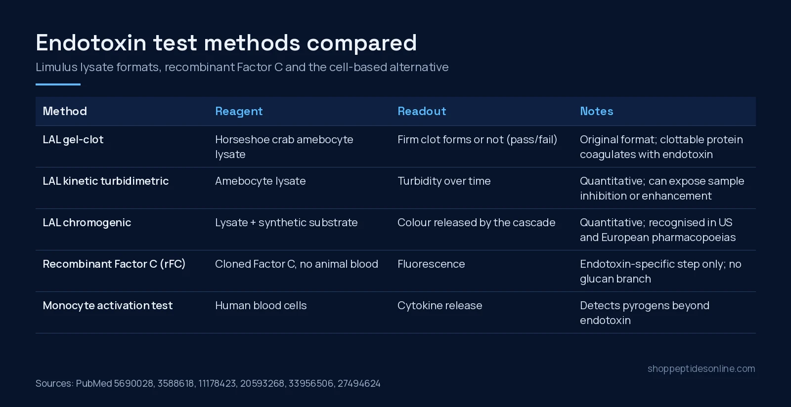
The test comes from the horseshoe crab. Its blood cells (amebocytes) contain a clottable protein that coagulates in the presence of endotoxin[4]. Biochemical work later mapped the cascade: several serine protease zymogens and the clottable protein coagulogen, triggered by bacterial endotoxin and also by (1,3)-beta-D-glucan[5]. Factor C is the endotoxin-sensing first step. In reconstitution experiments, purified Factor C, Factor B, proclotting enzyme and coagulogen together formed a gel in the presence of endotoxin[6].
LAL testing is now written into pharmacopoeias. Both the United States and European Pharmacopoeias describe the LAL bacterial endotoxin test, and endotoxin limits measured by LAL have replaced the older rabbit pyrogen test in many monographs[9].
Gel-clot, turbidimetric and chromogenic formats
- Gel-clot is the original readout: the lysate either forms a firm clot at a given sensitivity or it doesn’t, so the result is a pass/fail limit test[4][9].
- Kinetic turbidimetric assays track the rising cloudiness as the clot forms. Work on this format showed it can quantify endotoxin and expose a sample’s inhibiting or enhancing effects, which often resolve on dilution[7]. Kinetic analysis can also help separate a true endotoxin signal from a glucan signal[8].
- Chromogenic assays add a synthetic substrate that the cascade’s final enzyme cleaves to release colour. This led to kinetic quantitative chromogenic methods used to control endotoxin in pharmaceuticals and medical devices[9].
Recombinant Factor C: the animal-free option
rFC is Factor C produced by genetic engineering. The recombinant proenzyme is activated by trace endotoxin and underpins fluorescence-based endotoxin assays[10]. The motivation was twofold: variation in the sensitivity and specificity of LAL, and the declining supply of horseshoe crabs[10][14]. Because rFC uses only the Factor C step, it does not include the glucan-triggered branch of the natural lysate cascade[10][5].
The comparison data are broadly reassuring. A pharmaceutical validation reported an end-point fluorescence rFC method as equivalent or superior to the compendial endotoxin test[11]. A review of LAL-versus-rFC studies concluded the two are equivalent and comparable[12]. In four human vaccines with complex matrices, the rFC assays performed at least as well as the LAL assays[13]. A conservation-focused study likewise confirmed rFC’s efficacy as a synthetic alternative to horseshoe crab blood[14].
Side-by-side comparison
| Method | What reacts | Result type | Watch-outs |
|---|---|---|---|
| LAL gel-clot | Whole lysate cascade[4] | Pass/fail at a set sensitivity | Not quantitative; glucans can react[5] |
| LAL kinetic turbidimetric | Whole lysate cascade | Quantitative (turbidity over time)[7] | Sample inhibition or enhancement; check by dilution[7] |
| LAL chromogenic | Lysate plus synthetic colour substrate[9] | Quantitative (colour) | Same lysate-based interferences |
| Recombinant Factor C | Cloned Factor C only[10] | Quantitative (fluorescence)[11] | No glucan branch; masking (LER) can still hide endotoxin[16] |
| Monocyte activation test | Human blood cells[19] | Cytokine response | An in vitro pyrogen test that replaces the rabbit test; not endotoxin-specific |
Low endotoxin recovery: when the test misses it
From 2013, labs reported that endotoxin spiked into certain biological products could not be fully recovered by LAL over time. The phenomenon became known as low endotoxin recovery (LER), and FDA began requesting spike/hold recovery data in product applications[15]. In one study, chelators and detergents common in buffer formulations masked endotoxin. The masked LPS was not detected by Factor C-based assays but was still active in a TLR4 reporter assay and in primary human monocytes[16]. Reviews link LER to how formulation components disturb LPS aggregates, and the mechanism is still not fully settled[17][18].
The practical point for a lab: if your peptide is dissolved in a buffer with detergents or chelators, a “low” endotoxin result is more trustworthy when the test included a spike-recovery control in that same matrix.
What a typical peptide COA does and doesn’t cover
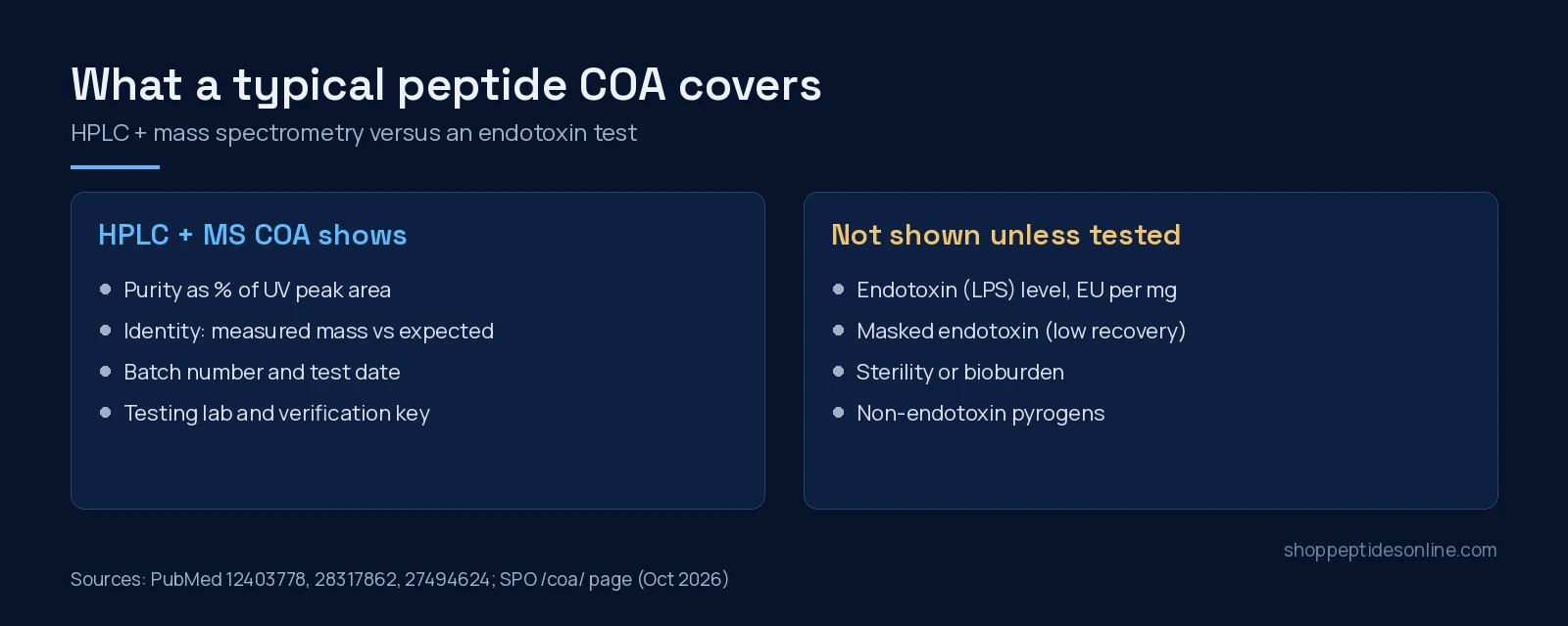
Most research-peptide certificates of analysis report two things: HPLC purity (the share of UV peak area belonging to the target peptide) and mass-spectrometry identity (whether the measured mass matches the expected sequence). Our guide to HPLC purity vs mass spectrometry identity explains both. Neither measures endotoxin. A peptide can pass at high purity and correct mass while still carrying LPS at levels that change a cell assay, as the Hsp70 example shows for a protein preparation[3].
Where Shop Peptides Online stands: our COA page lists third-party reports with product identity, batch, reported purity, test date and the testing lab. At the time of writing it does not list endotoxin results, so do not assume a batch has been endotoxin-tested unless its own report says so. If your protocol needs a documented endotoxin level, plan to test the material in your own lab or through an accredited testing service, using a method validated for your sample matrix.
Diluent choice matters too. Our comparison of bacteriostatic water vs sterile water covers what each is and isn’t designed for. When you’re ready to source material, you can buy peptides online in Canada from our research catalogue and review each batch’s certificate first.
What the evidence does not show
- There is no published data here on endotoxin levels in commercial research peptides as a category. The contamination examples above involve recombinant proteins[2][3], not synthetic peptides.
- rFC-versus-LAL equivalence has been shown in specific products and matrices[11][13]. Any method still needs validating in your own sample matrix.
- Masking can hide biologically active endotoxin from both LAL and rFC[16], so a negative result is only as good as its recovery controls.
- Endotoxin tests detect endotoxin only. Other pyrogens need a broader assay such as the monocyte activation test[19].
Frequently asked questions
Does a high HPLC purity result mean a peptide is endotoxin-free?
No. HPLC purity is the share of UV peak area belonging to the target peptide, and mass spectrometry confirms its mass. Neither measures bacterial lipopolysaccharide. In one study, endotoxin present at well under one part per thousand by mass was enough to drive a cytokine response in macrophages.
What is the difference between LAL and rFC?
LAL is a lysate of horseshoe crab blood cells containing a whole clotting cascade. rFC is a cloned version of Factor C, the cascade’s endotoxin sensor, produced without animal blood. Comparison studies have reported rFC to be equivalent or comparable to LAL for endotoxin detection.
What is low endotoxin recovery?
Low endotoxin recovery (LER) is the failure of Limulus-based tests to detect endotoxin that was spiked into some formulations, usually those containing chelators or detergents. In one study the masked endotoxin was still active in cell-based assays.
Why can glucans interfere with LAL tests?
The lysate cascade can be triggered by both endotoxin and (1,3)-beta-D-glucan, so glucan-containing samples can react. Kinetic analysis can help tell them apart, and rFC uses only the endotoxin-sensing Factor C step.
Does Shop Peptides Online test for endotoxin?
Our COA page lists third-party reports with identity, batch, purity, test date and testing lab. It does not currently list endotoxin results, so do not assume a batch has been endotoxin-tested unless its own report says so.
References
- Poltorak A, He X, Smirnova I et al. (1998). Defective LPS signaling in C3H/HeJ and C57BL/10ScCr mice: mutations in Tlr4 gene. Science. PMID: 9851930. doi: 10.1126/science.282.5396.2085.
- Wakelin SJ, Sabroe I, Gregory CD et al. (2006). “Dirty little secrets”–endotoxin contamination of recombinant proteins. Immunol Lett. PMID: 16765451. doi: 10.1016/j.imlet.2006.04.007.
- Gao B, Tsan MF (2003). Endotoxin contamination in recombinant human heat shock protein 70 (Hsp70) preparation is responsible for the induction of tumor necrosis factor alpha release by murine macrophages. J Biol Chem. PMID: 12403778. doi: 10.1074/jbc.M208742200.
- Levin J, Bang FB (1968). Clottable protein in Limulus; its localization and kinetics of its coagulation by endotoxin. Thromb Diath Haemorrh. PMID: 5690028.
- Iwanaga S (1993). The limulus clotting reaction. Curr Opin Immunol. PMID: 8452677. doi: 10.1016/0952-7915(93)90084-6.
- Iwanaga S (2007). Biochemical principle of Limulus test for detecting bacterial endotoxins. Proc Jpn Acad Ser B Phys Biol Sci. PMID: 24019589. doi: 10.2183/pjab.83.110.
- Remillard JF, Gould MC, Roslansky PF et al. (1987). Quantitation of endotoxin in products using the LAL kinetic turbidimetric assay. Prog Clin Biol Res. PMID: 3588618.
- Tsuchiya M, Oishi H, Takaoka A et al. (1990). Discrimination between endotoxin and (1—-3)-beta-D-glucan using turbidimetric kinetic assay with Limulus amebocyte lysate. Chem Pharm Bull (Tokyo). PMID: 2285986. doi: 10.1248/cpb.38.2523.
- Berzofsky RN (1995). Endotoxin Detection in Pharmaceuticals and Medical Devices with Kinetic-QCL, a Kinetic-Quantitative Chromogenic Limulus Amebocyte Lysate Assay. ALTEX. PMID: 11178423.
- Ding JL, Ho B (2010). Endotoxin detection–from limulus amebocyte lysate to recombinant factor C. Subcell Biochem. PMID: 20593268. doi: 10.1007/978-90-481-9078-2_9.
- Bolden J, Smith K (2017). Application of Recombinant Factor C Reagent for the Detection of Bacterial Endotoxins in Pharmaceutical Products. PDA J Pharm Sci Technol. PMID: 28733334. doi: 10.5731/pdajpst.2017.007849.
- Tindall B, Demircioglu D, Uhlig T (2021). Recombinant bacterial endotoxin testing: a proven solution. Biotechniques. PMID: 33956506. doi: 10.2144/btn-2020-0165.
- Marius M, Vacher F, Bonnevay T (2020). Comparison of Limulus Amoebocyte Lysate and Recombinant Factor C Assays for Endotoxin Detection in Four Human Vaccines with Complex Matrices. PDA J Pharm Sci Technol. PMID: 32179709. doi: 10.5731/pdajpst.2019.010389.
- Maloney T, Phelan R, Simmons N (2018). Saving the horseshoe crab: A synthetic alternative to horseshoe crab blood for endotoxin detection. PLoS Biol. PMID: 30312293. doi: 10.1371/journal.pbio.2006607.
- Bolden JS, Warburton RE, Phelan R et al. (2016). Endotoxin recovery using limulus amebocyte lysate (LAL) assay. Biologicals. PMID: 27470947. doi: 10.1016/j.biologicals.2016.04.009.
- Schwarz H, Gornicec J, Neuper T et al. (2017). Biological Activity of Masked Endotoxin. Sci Rep. PMID: 28317862. doi: 10.1038/srep44750.
- Cao Y, Zhang Y, Qiu F (2021). Low endotoxin recovery and its impact on endotoxin detection. Biopolymers. PMID: 34407207. doi: 10.1002/bip.23470.
- Gorman A, Golovanov AP (2022). Lipopolysaccharide Structure and the Phenomenon of Low Endotoxin Recovery. Eur J Pharm Biopharm. PMID: 36272656. doi: 10.1016/j.ejpb.2022.10.006.
- Fennrich S, Hennig U, Toliashvili L et al. (2016). More than 70 years of pyrogen detection: Current state and future perspectives. Altern Lab Anim. PMID: 27494624. doi: 10.1177/026119291604400305.
