For laboratory research only. Not for human or animal use. This article gives no dosing, administration or therapeutic advice.
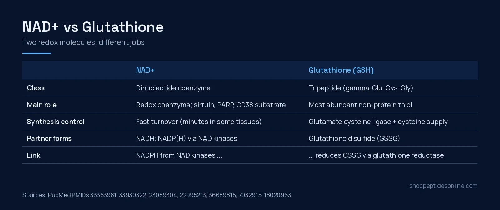
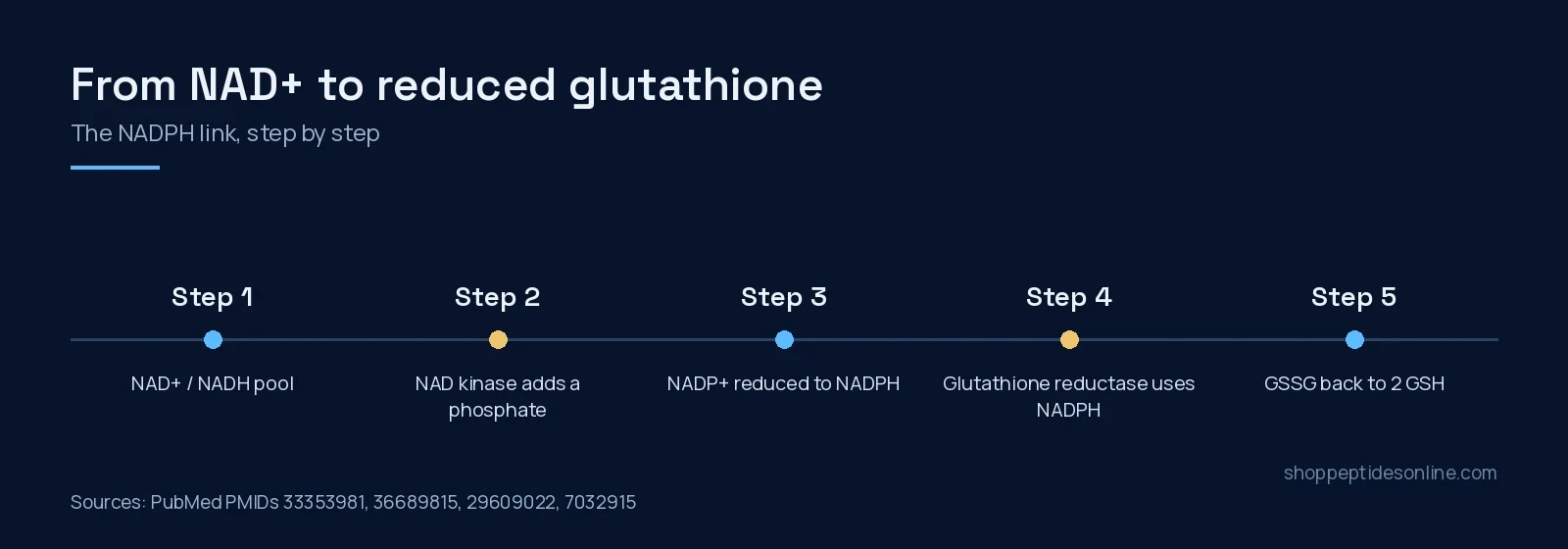
Short answer: NAD+ and glutathione are both central to cellular redox chemistry, but they do different jobs. NAD+ is a dinucleotide coenzyme that carries electrons in energy metabolism and is also consumed as a substrate by sirtuins, PARPs and CD38[1]. Glutathione (GSH) is a tripeptide, gamma-glutamyl-cysteinyl-glycine, and the most abundant non-protein thiol in mammalian tissues, where it defends against oxidative stress[2][3]. The two meet through NADPH: NAD kinases phosphorylate NAD(H) to make NADP(H)[4], and glutathione reductase uses NADPH to turn oxidized glutathione disulfide back into GSH[5].
Side by side
| NAD+ | Glutathione (GSH) | |
|---|---|---|
| Chemical class | Dinucleotide coenzyme (not a peptide)[1] | Tripeptide, gamma-glutamyl-cysteinyl-glycine[2] |
| Main role | Redox coenzyme in energy metabolism; substrate for sirtuins, CD38 and PARPs[1] | Thiol antioxidant; also redox signalling and xenobiotic detoxification[3] |
| Made from | NAD pathway precursors; turned over with a half-life of minutes in some tissues[6] | Cysteine, glutamate and glycine via glutamate cysteine ligase then GSH synthetase[3] |
| Rate-limiting step | Not a single step; synthesis and consumption are both dynamic[6] | Glutamate cysteine ligase and cysteine supply[3] |
| Oxidized form | NADH is the reduced partner; NADP(H) is a phosphorylated relative[7] | Glutathione disulfide, reduced back by glutathione reductase with NADPH[5] |
| Where it is made | Different compartments need their own NADP-making enzymes, since NADP cannot cross membranes[4] | In the cytosol, under tight regulation[3] |
| At SPO | NAD+ 1000mg | Not sold |
Two different molecules
NAD+ is a coenzyme for oxidation–reduction reactions, which makes it central to energy metabolism. It is also an essential cofactor for enzymes that do not use it for redox chemistry: sirtuins, CD38 and poly(ADP-ribose) polymerases. Through those enzymes it touches DNA repair, chromatin remodelling, senescence and immune cell function[1]. A 2021 perspective adds that NAD is turned over quickly, with a half-life on the order of minutes in some tissues, and that how NAD and its precursors enter cells and organelles is still an open question[6].
Glutathione is built in the cytosol in two enzymatic steps. Glutamate cysteine ligase, with catalytic and modifier subunits, is rate-limiting, and cysteine availability is the other main control point; GSH synthetase completes the tripeptide. Nrf2, AP-1 and NF-kappaB regulate the genes for both enzymes[3]. Beyond antioxidant defence, GSH is a determinant of redox signalling and is involved in detoxifying foreign compounds[3].
The link: NADP(H)
A 2008 review groups NAD and NADP as common mediators of energy metabolism, mitochondrial function, antioxidation, calcium signalling and cell death, and identifies NADPH as a key component of cellular antioxidant systems[7]. NADP is made from NAD by NAD kinases. Because NADP cannot cross subcellular membranes, cells keep separate NAD kinases in different compartments; the mitochondrial enzyme NADK2 phosphorylates NAD(H) to produce mitochondrial NADP(H)[4].
Glutathione reductase is the enzyme that closes the loop. Purified from human red cells, it is a flavoenzyme that reduces glutathione disulfide using NADPH[5]. Mutagenesis of its NADPH-binding motif in E. coli showed how tightly that coenzyme site is tuned: most substitutions at one conserved glycine virtually inactivated the enzyme[8]. More broadly, NADPH supplies the reducing power for the cell’s NADPH-dependent disulfide reductase systems[9].
Where they show up together in data
In human pelvic skin samples from 49 donors, tissue NAD+ fell with age in both sexes. In males, PARP activity rose with age and correlated inversely with NAD+, and lipid oxidation rose with age; several of these associations were weaker in females[10]. That is a correlational study, not a test of either molecule as an intervention, but it shows why oxidative-stress markers and NAD+ are often measured in the same experiment.
Which one fits which question?
- Sirtuin, PARP or CD38 activity, energy metabolism, NAD turnover: NAD+[1][6].
- Thiol redox state, GSH synthesis regulation, detoxification pathways: glutathione and its synthetic enzymes[3].
- How energy metabolism feeds antioxidant capacity: the NAD kinase to NADPH to glutathione reductase axis[4][5].
For NAD+ precursors, see NAD+ vs NMN vs NR, and our NAD+ research page covers the product. For the full catalogue, see our guide to buy peptides online in Canada.
Frequently asked questions
Is NAD+ a peptide like glutathione?
No. NAD+ is a dinucleotide coenzyme. Glutathione is a tripeptide made of glutamate, cysteine and glycine.
How are NAD+ and glutathione connected?
NAD kinases convert NAD(H) into NADP(H), and glutathione reductase uses NADPH to reduce glutathione disulfide back to glutathione.
What limits glutathione synthesis in cells?
The rate-limiting enzyme glutamate cysteine ligase and the availability of cysteine.
Why measure NADPH alongside glutathione?
Glutathione reductase uses NADPH to regenerate reduced glutathione from glutathione disulfide, so NADPH supply is one input to the reduced glutathione pool.
Does Shop Peptides Online sell glutathione?
No. SPO lists NAD+ for laboratory research only. Glutathione is not part of the catalogue.
References
- Covarrubias AJ, Perrone R, Grozio A et al. (2021). NAD(+) metabolism and its roles in cellular processes during ageing. Nat Rev Mol Cell Biol. PMID: 33353981. doi: 10.1038/s41580-020-00313-x.
- Morris D, Khurasany M, Nguyen T et al. (2013). Glutathione and infection. Biochim Biophys Acta. PMID: 23089304. doi: 10.1016/j.bbagen.2012.10.012.
- Lu SC (2013). Glutathione synthesis. Biochim Biophys Acta. PMID: 22995213. doi: 10.1016/j.bbagen.2012.09.008.
- Zhang R, Zhang K (2023). Mitochondrial NAD kinase in health and disease. Redox Biol. PMID: 36689815. doi: 10.1016/j.redox.2023.102613.
- Untucht-Grau R, Schirmer RH, Schirmer I et al. (1981). Glutathione reductase from human erythrocytes: amino-acid sequence of the structurally known FAD-binding domain. Eur J Biochem. PMID: 7032915. doi: 10.1111/j.1432-1033.1981.tb05718.x.
- Chini CCS, Zeidler JD, Kashyap S et al. (2021). Evolving concepts in NAD(+) metabolism. Cell Metab. PMID: 33930322. doi: 10.1016/j.cmet.2021.04.003.
- Ying W (2008). NAD+/NADH and NADP+/NADPH in cellular functions and cell death: regulation and biological consequences. Antioxid Redox Signal. PMID: 18020963. doi: 10.1089/ars.2007.1672.
- Rescigno M, Perham RN (1994). Structure of the NADPH-binding motif of glutathione reductase: efficiency determined by evolution. Biochemistry. PMID: 8180198. doi: 10.1021/bi00185a008.
- Miller CG, Holmgren A, Arnér ESJ et al. (2018). NADPH-dependent and -independent disulfide reductase systems. Free Radic Biol Med. PMID: 29609022. doi: 10.1016/j.freeradbiomed.2018.03.051.
- Massudi H, Grant R, Braidy N et al. (2012). Age-associated changes in oxidative stress and NAD+ metabolism in human tissue. PLoS One. PMID: 22848760. doi: 10.1371/journal.pone.0042357.
- Buonvicino D, Ranieri G, Pittelli M et al. (2021). SIRT1-dependent restoration of NAD+ homeostasis after increased extracellular NAD+ exposure. J Biol Chem. PMID: 34097876. doi: 10.1016/j.jbc.2021.100855.