For laboratory research only. Not for human or animal use. This article gives no dosing, administration or therapeutic advice.
Short answer: thymosin beta-4 (Tβ4) is a 43-amino-acid protein that cells make naturally. “TB-500” is a trade name, and when doping-control chemists analysed products sold under it, they found a synthetic seven-residue piece of that protein, Ac-LKKTETQ (residues 17 to 23).[1],[2] The two differ more than five-fold in mass, have different research records, and only the full protein has been tested in human trials. Because vendors use “TB-500” for both, the only reliable way to know which one a vial holds is the mass on its certificate of analysis.
Two molecules, one name
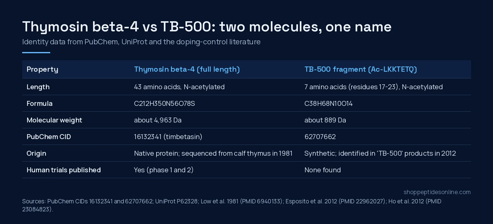
Thymosin beta-4 was first sequenced from calf thymus in 1981: 43 residues, an acetylated N-terminus and an isoelectric point of 5.1.[3] It turned out to be the main G-actin-sequestering protein in eukaryotic cells, which is why it appears in so many tissues.[4] PubChem lists the full-length molecule (INN timbetasin) at C212H350N56O78S and a molecular weight of about 4,963 Da.
The fragment is a different compound with its own PubChem record (CID 62707662): C38H68N10O14, about 889 Da, synonym “TB-500”.[1] Equine doping-control chemists described the “key ingredient” of a veterinary TB-500 preparation as N-acetylated LKKTETQ, and built a method to detect it and its metabolites in horse urine and plasma.[2] In other words, the people with the most reason to know exactly what TB-500 is (anti-doping labs) found a fragment, not the whole protein.
| Thymosin beta-4 | TB-500 fragment | |
|---|---|---|
| Sequence | Ac-SDKPDMAEIEKFDKSKLKKTETQEKNPLPSKETIEQEKQAGES (43 aa) | Ac-LKKTETQ (7 aa, residues 17-23) |
| Molecular weight | about 4,963 Da (PubChem CID 16132341) | about 889 Da (PubChem CID 62707662) |
| Where it comes from | Native protein, first sequenced from calf thymus[3] | Synthetic; identified in TB-500 products[1],[2] |
| Main research models | Cell culture, rat, mouse, and small human trials | Cell and chick aortic-ring assays; doping-control analysis |
| Mass a COA should report | near 4,963 Da | near 889 Da |
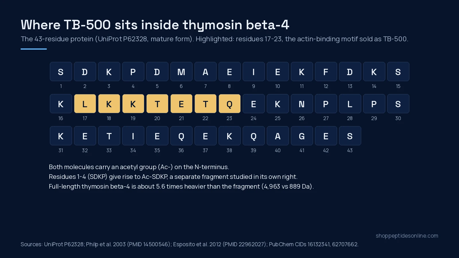
The sequence map below shows where the fragment sits: the actin-binding stretch in the first half of the protein. The first four residues, SDKP, are the source of a third molecule, Ac-SDKP, which researchers study separately.[5],[6]
Why the fragment was made
The case for the short peptide comes mostly from one 2003 study. Using natural Tβ4, proteolytic fragments and synthetic peptides, the authors found that the seven-amino-acid actin-binding motif was essential for Tβ4’s angiogenic activity. In human umbilical vein endothelial cell migration assays and chick aortic-arch sprouting assays, the full protein and the motif showed near-identical activity, and peptides missing any part of the motif were inactive.[7]
That is a real finding, but it is narrow. It covers endothelial migration, adhesion and sprouting in cell and explant models. It does not show that the fragment reproduces everything the 43-residue protein does elsewhere, such as the PINCH and integrin-linked kinase interaction reported in mouse heart studies.[8]
What has been studied for each, by model
Most published biology is on full-length Tβ4. Papers on “TB-500” are rarer, and some do not say which sequence they used.
| Study | Molecule | Model | What was reported |
|---|---|---|---|
| Malinda 1999[9] | Tβ4 | Rat full-thickness wound; keratinocyte migration assay | Faster re-epithelialisation and more collagen deposition than saline controls |
| Philp 2003[7] | Tβ4 and the LKKTETQ motif | Human endothelial cells; chick aortic arches | Near-identical activity for protein and motif; motif required |
| Bock-Marquette 2004[8] | Tβ4 | Mouse coronary-ligation model; cultured cardiomyocytes | Complex with PINCH and ILK, Akt activation, better early myocyte survival |
| Smart 2007[5] | Tβ4 and Ac-SDKP | Mouse heart and epicardial explants | Tβ4 needed for coronary vessel development; Ac-SDKP could not rescue Tβ4-deficient hearts |
| Spurney 2010[10] | Tβ4 | mdx mouse (muscular dystrophy model) | More regenerating fibres, but no improvement in grip strength, cardiac function or fibrosis |
| Conte 2015[6] | Tβ4 and Ac-SDKP | Human lung fibroblasts; bleomycin mouse | Tβ4 failed to prevent fibrosis at 14 and 21 days; Ac-SDKP was the more active anti-fibrotic in vitro |
| Biçer 2026[11] | “Synthetic thymosin beta-4 (TB-500)” | Rat Achilles tendon repair | Better histology and load to failure versus controls; no added effect when combined with BPC-157; sequence not stated in the abstract |
Two rows deserve attention. The muscular dystrophy and lung fibrosis studies are negative on their main functional outcomes, which matters because summaries of Tβ4 research tend to leave them out.[10],[6] And the 2026 tendon paper shows the naming problem inside the literature itself: its abstract calls the test article “synthetic thymosin beta-4 (TB-500)” without giving a sequence, so a reader cannot tell which molecule was used.[11]
Human data exist only for the full protein
Full-length Tβ4 has gone through investigational trials. A first-in-human phase 1 study of a recombinant form enrolled 54 healthy volunteers in single ascending-dose cohorts and 30 in multiple-dose cohorts; adverse events were mild to moderate, and there were no dose-limiting toxicities or serious adverse events.[12] A randomised, double-masked phase 2 trial in severe dry eye had nine participants, which its authors call small.[13] A European placebo-controlled study in venous ulcers published its design and early enrolment in 2007.[14]
None of that transfers to the fragment. We found no published human trial of Ac-LKKTETQ. Its human-relevant literature is doping control: detection methods, metabolites and the description of TB-500 as a product “suspected to possess doping potential.”[1],[2] Neither molecule is an approved medicine, and both are supplied here only for in-vitro and laboratory research.
Reading the mass on a COA
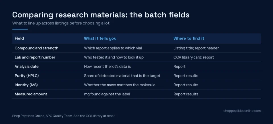
This is where the difference becomes practical. An HPLC purity figure tells you what share of the sample sits in the main peak. It does not say what that peak is. Identity comes from mass spectrometry, and here the two candidates are easy to separate: roughly 4,963 Da for the full protein against roughly 889 Da for the fragment. Esposito and colleagues used high-resolution LC-MS for exactly this purpose.[1]
SPO’s own Janoshik reports show why the label alone is not enough. The report for the TB-500 5mg listing (#219841, analysed 20 August 2026) prints its result as “TB-500 (TB-4)”. The KLOW blend report (#172068) prints “TB-500 (889 Da)”, the fragment’s mass. Same trade name, two readings. When you compare TB-500 listings, check which molecule the report names, then open the report itself in the COA library.
The TB-500 research hub carries the full study table and storage notes. The individual listings are TB-500 5mg and TB-500 10mg. For how SPO ships and documents orders across Canada, see our page on how to buy peptides online in Canada.
Frequently asked questions
Is TB-500 the same thing as thymosin beta-4?
Not necessarily. Thymosin beta-4 is a 43-amino-acid protein of about 4,963 Da. Doping-control laboratories that analysed products sold as TB-500 found the seven-residue fragment Ac-LKKTETQ (about 889 Da), which is residues 17 to 23 of the protein. The name is used for both, so the molecule has to be confirmed by mass.
Which of the two has been tested in people?
Only full-length thymosin beta-4. Published work includes a phase 1 safety and pharmacokinetics study of a recombinant form in healthy volunteers and small phase 2 studies in dry eye and venous ulcers. We found no published human trial of the Ac-LKKTETQ fragment.
How can a lab tell which molecule is in a vial?
By mass spectrometry. An identity result near 4,963 Da points to full-length thymosin beta-4; a result near 889 Da points to the fragment. HPLC purity alone cannot tell them apart, because it reports the share of the main peak, not what that peak is.
Why would anyone study the short fragment instead of the protein?
In endothelial cell and chick aortic-arch assays, the seven-residue actin-binding motif matched the full protein’s activity on cell migration and vessel sprouting, while peptides lacking the motif were inactive. That 2003 result is the main reason the fragment exists as a research tool. It has not been tested in human studies.
Related reading: BPC-157 Canada: 5mg and 10mg research vials, batch COA and shipping across provinces.
References
- Esposito S, Deventer K, Goeman J et al. (2012). Synthesis and characterization of the N-terminal acetylated 17-23 fragment of thymosin beta 4 identified in TB-500, a product suspected to possess doping potential. Drug Test Anal. PMID: 22962027. doi: 10.1002/dta.1402.
- Ho EN, Kwok WH, Lau MY et al. (2012). Doping control analysis of TB-500, a synthetic version of an active region of thymosin β₄, in equine urine and plasma by liquid chromatography-mass spectrometry. J Chromatogr A. PMID: 23084823. doi: 10.1016/j.chroma.2012.09.043.
- Low TL, Hu SK, Goldstein AL (1981). Complete amino acid sequence of bovine thymosin beta 4: a thymic hormone that induces terminal deoxynucleotidyl transferase activity in thymocyte populations. Proc Natl Acad Sci U S A. PMID: 6940133. doi: 10.1073/pnas.78.2.1162.
- Goldstein AL, Hannappel E, Kleinman HK (2005). Thymosin beta4: actin-sequestering protein moonlights to repair injured tissues. Trends Mol Med. PMID: 16099219. doi: 10.1016/j.molmed.2005.07.004.
- Smart N, Risebro CA, Melville AA et al. (2007). Thymosin beta4 induces adult epicardial progenitor mobilization and neovascularization. Nature. PMID: 17108969. doi: 10.1038/nature05383.
- Conte E, Iemmolo M, Fruciano M et al. (2015). Effects of thymosin β4 and its N-terminal fragment Ac-SDKP on TGF-β-treated human lung fibroblasts and in the mouse model of bleomycin-induced lung fibrosis. Expert Opin Biol Ther. PMID: 26098610. doi: 10.1517/14712598.2015.1026804.
- Philp D, Huff T, Gho YS et al. (2003). The actin binding site on thymosin beta4 promotes angiogenesis. FASEB J. PMID: 14500546. doi: 10.1096/fj.03-0121fje.
- Bock-Marquette I, Saxena A, White MD et al. (2004). Thymosin beta4 activates integrin-linked kinase and promotes cardiac cell migration, survival and cardiac repair. Nature. PMID: 15565145. doi: 10.1038/nature03000.
- Malinda KM, Sidhu GS, Mani H et al. (1999). Thymosin beta4 accelerates wound healing. J Invest Dermatol. PMID: 10469335. doi: 10.1046/j.1523-1747.1999.00708.x.
- Spurney CF, Cha HJ, Sali A et al. (2010). Evaluation of skeletal and cardiac muscle function after chronic administration of thymosin beta-4 in the dystrophin deficient mouse. PLoS One. PMID: 20126456. doi: 10.1371/journal.pone.0008976.
- Biçer O, Adanir O, Güleryüz Y et al. (2026). Effects of BPC-157 and TB-500 on Achilles tendon healing in rats: A histopathological and biomechanical study. Jt Dis Relat Surg. PMID: 42542926. doi: 10.52312/jdrs.2026.2951.
- Wang X, Liu L, Qi L et al. (2021). A first-in-human, randomized, double-blind, single- and multiple-dose, phase I study of recombinant human thymosin β4 in healthy Chinese volunteers. J Cell Mol Med. PMID: 34346165. doi: 10.1111/jcmm.16693.
- Sosne G, Dunn SP, Kim C (2015). Thymosin β4 significantly improves signs and symptoms of severe dry eye in a phase 2 randomized trial. Cornea. PMID: 25826322. doi: 10.1097/ICO.0000000000000379.
- Guarnera G, DE Rosa A, Camerini R (2007). Thymosin beta-4 and venous ulcers: clinical remarks on a European prospective, randomized study on safety, tolerability, and enhancement on healing. Ann N Y Acad Sci. PMID: 17495250. doi: 10.1196/annals.1415.003.