For laboratory research only. Not for human or animal use. This article gives no dosing, administration or therapeutic advice.
Short answer: a synthetic peptide is a salt. The peptide carries positive charges, and a counter-ion balances them. After standard solid-phase synthesis and HPLC purification that counter-ion is usually trifluoroacetate (TFA), because TFA is the cleavage agent and the ion-pairing reagent[1]. Acetate and hydrochloride are the usual alternatives. The choice rarely matters for identity or HPLC purity, but published studies show it can matter for cell assays, animal models, infrared spectroscopy and the true peptide content of a vial.
Side by side
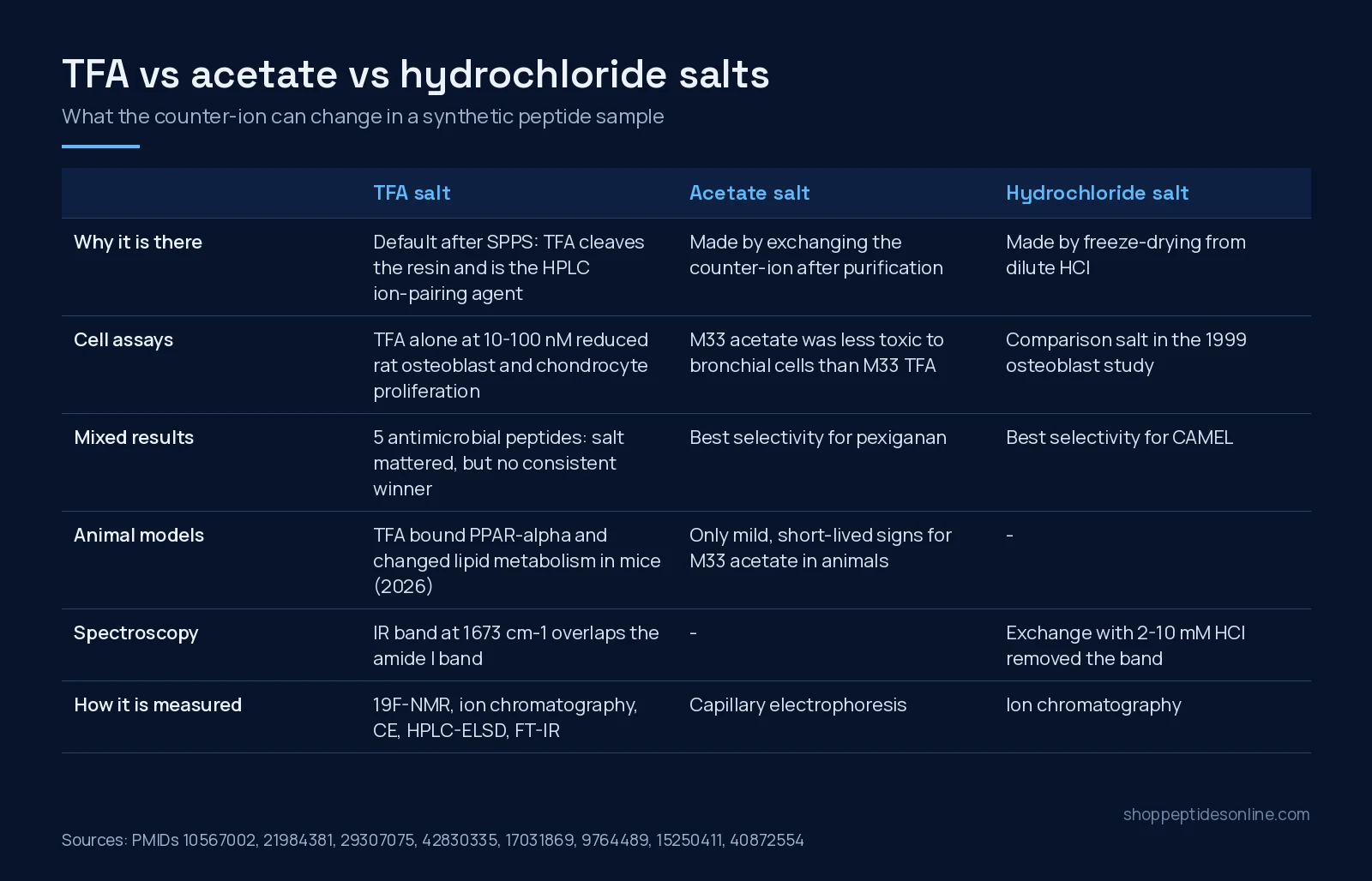
| TFA salt | Acetate salt | Hydrochloride salt | |
|---|---|---|---|
| How it arises | Default after SPPS cleavage and RP-HPLC with TFA[1][6] | Counter-ion exchange after purification | Freeze-drying from dilute HCl[7][1] |
| Cell-assay record | TFA alone at 10–100 nM reduced osteoblast and chondrocyte proliferation[2] | M33 acetate 5–30% less toxic than M33 TFA in bronchial cell lines[4] | Reference salt for the 1999 osteoblast comparison[2] |
| Animal-model record | TFA bound PPAR-alpha and altered lipid metabolism in three mouse strains[3] | M33 acetate gave only mild, short-lived signs where the TFA salt gave manifest toxicity[4] | Not compared in these sources |
| Infrared spectroscopy | Band at 1673 cm−1 overlaps or hides the amide I band[7] | Not reported as interfering in these sources | Used to remove the TFA band[7] |
| Status in drug development | Most marketed peptide drugs are hydrochloride or acetate salts; regulators view TFA salts less favourably[6] | ||
Where the TFA comes from
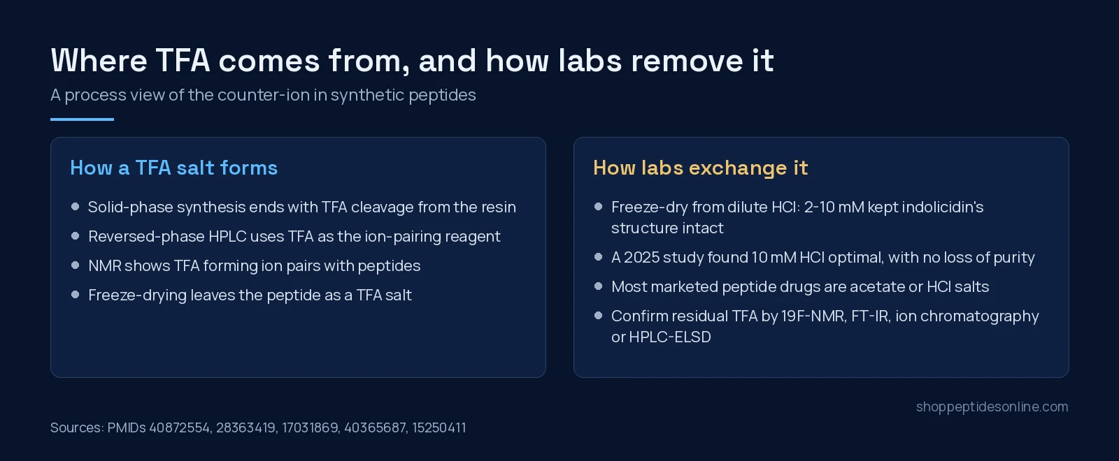
Solid-phase peptide synthesis builds the chain on a resin, then cleaves it with strong acid, usually TFA. Purification by reversed-phase HPLC then runs in acetonitrile and water with TFA as the ion-pairing reagent[1]. This is not a loose analogy: fluorine-proton NMR experiments gave direct evidence of ion pairs forming between a peptide and TFA in acetonitrile-water[9]. When the purified fractions are freeze-dried, the peptide comes out as its TFA salt. Peptides with more basic residues (lysine, arginine, histidine) carry more positive charges and so can carry more counter-ion.
The counter-ion is not fixed forever. In a study of the caveolin-1 scaffolding peptide CSP7, the formulation and process changed how much counter-ion stayed with the peptide, and the pH modifier had the largest effect[14].
Why researchers care: the published effects
Cell proliferation. The best-known paper is a 1999 study in fetal rat osteoblasts. TFA at 10−8 to 10−7 M reduced cell numbers and thymidine incorporation after 24 hours, with similar effects in articular chondrocytes and neonatal mouse calvariae. When TFA and hydrochloride salts of amylin, amylin-(1-8) and calcitonin were compared, the TFA salts consistently gave less proliferation, enough to hide a proliferative effect or to suggest an antiproliferative one that wasn’t the peptide’s own[2]. The authors concluded that this is likely relevant to peptide studies above 10−9 M.
Toxicity comparisons. For the branched antimicrobial peptide M33, the TFA and acetate salts did not differ substantially in antimicrobial efficacy in vitro or in vivo. The TFA salt was 5–30% more toxic to bronchial-derived cell lines, and produced clear toxicity signs in animals, where the acetate salt produced only mild signs that resolved within hours[4].
No universal winner. Five antimicrobial peptides (CAMEL, citropin 1.1, LL-37, pexiganan and temporin A) were each prepared as acetate, hydrochloride and TFA salts. The salt form made a substantial difference, but the pattern was not consistent between peptides. The best selectivity indexes belonged to CAMEL hydrochloride, pexiganan acetate and temporin A TFA[5]. In a peptide hydrogel platform, TFA and HCl salts were compared for biostability, cytotoxicity, release and rheology[6].
TFA as an active molecule. A 2026 study challenged the assumption that TFA is inert. In three mouse strains and in cultured liver cells, TFA induced peroxisome proliferation and changed lipid metabolism, both alone and as the counter-ion of several unrelated short peptides. The mechanistic evidence pointed to TFA binding PPAR-alpha[3]. The authors called for assessing TFA’s contribution to effects seen in studies using synthetic peptides. These are mouse and cell results.
Structural work. TFA’s infrared band at 1673 cm−1 overlaps the amide I band used to read secondary structure[7]. In a centrin-melittin model complex, TFA present as an excipient was associated with thermally induced aggregation of melittin[8].
How labs measure and remove it
An HPLC purity figure compares the target peptide with peptide-related impurities. The counter-ion is measured separately. In one certified-reference-material project, TFA was quantified by validated 19F-qNMR and treated as part of the mass balance, at nearly 25% of the material by mass[10]. Other validated methods include:
- Ion chromatography with suppressed conductivity, with detection limits below 90 ng/mL in buffer, protein and peptide samples[11].
- Capillary electrophoresis for both acetic acid and TFA in synthetic peptide samples[12].
- FT-IR, 19F-NMR and HPLC with an evaporative light-scattering detector, all validated to ICH guidelines in a 2025 study[1].
- Combined 1H-19F NMR, a non-destructive method that quantifies TFA without weighing the sample[13].
To remove TFA, the usual method is freeze-drying from dilute HCl. With indolicidin, 2–10 mM HCl removed essentially all TFA without changing secondary structure, while concentrations above 10 mM (unbuffered) could alter the structure and thermal stability[7]. The 2025 study found 10 mM HCl optimal, with no effect on peptide purity at any concentration tested, and found that membrane permeability depended on both sequence and salt form[1].
What this means when you order
- Identity and purity come first. Our COA library lists each batch’s identity, reported purity, test date and lab reference. HPLC purity vs mass spectrometry identity explains what those two tests cover.
- Salt form is a separate question. If your assay is sensitive to the counter-ion, such as cell proliferation, structural spectroscopy or metabolic read-outs, plan a counter-ion measurement, an exchange step or a vehicle control that matches it.
- Mass isn’t all peptide. Counter-ions and water add weight. Why a vial can test below its label covers measured content vs label.
- Handling still matters. Lyophilized vs reconstituted peptides covers storage of the powder and of solutions.
To browse the catalogue, see our research peptides or the guide to buy peptides online in Canada.
Frequently asked questions
What is a peptide counter-ion?
Most research peptides carry positive charges on basic residues and the N-terminus. A negatively charged ion, the counter-ion, balances them in the solid powder. In peptides made by solid-phase synthesis and purified by reversed-phase HPLC, that ion is usually trifluoroacetate (TFA), because TFA is used both to cleave the peptide from the resin and as the ion-pairing reagent during purification.
Is a TFA salt a purity problem?
Not in the HPLC sense. HPLC purity compares the target peptide with peptide-related impurities. The counter-ion is part of the salt and is measured separately, for example by 19F-NMR, ion chromatography or HPLC with an evaporative light-scattering detector. A sample can be high-purity by HPLC and still be a TFA salt.
Can TFA affect cell-culture results?
In published cell studies, yes. TFA alone at 10 to 100 nM reduced proliferation of fetal rat osteoblasts and articular chondrocytes, and TFA salts of amylin and calcitonin gave less proliferation than their hydrochloride salts. A 2026 mouse study found TFA itself bound PPAR-alpha and changed lipid metabolism. Effects depend on the peptide, the cell type and the concentration.
Is acetate always better than TFA?
No single salt wins in every model. Across five antimicrobial peptides tested as acetate, hydrochloride and TFA salts, the salt form changed activity and cytotoxicity, but the pattern was not consistent from peptide to peptide. Most marketed peptide drugs are acetate or hydrochloride salts, and regulators view TFA salts less favourably.
How do labs remove TFA from a peptide?
The common route is repeated freeze-drying from dilute hydrochloric acid, which swaps TFA for chloride. One study found 2 to 10 mM HCl removed essentially all TFA from indolicidin without changing its secondary structure, while higher concentrations could alter it. A 2025 study validated 10 mM HCl as optimal with no loss of purity. Ion exchange to acetate is the other common option.
Does the salt form change how much peptide is in a vial?
It can. The counter-ion adds mass without adding peptide. In one peptide reference material, TFA made up nearly 25% of the mass. That is one reason measured peptide content and label weight can differ.
References
- Erckes V, Streuli A, Chamera Rendueles L et al. (2025). Towards a Consensus for the Analysis and Exchange of TFA as a Counterion in Synthetic Peptides and Its Influence on Membrane Permeation. Pharmaceuticals (Basel). PMID: 40872554. doi: 10.3390/ph18081163.
- Cornish J, Callon KE, Lin CQ et al. (1999). Trifluoroacetate, a contaminant in purified proteins, inhibits proliferation of osteoblasts and chondrocytes. Am J Physiol. PMID: 10567002. doi: 10.1152/ajpendo.1999.277.5.E779.
- Tang W, Black AS, Moench R et al. (2026). Trifluoroacetate mediated reduction of atherosclerosis in mice. Nat Commun. PMID: 42830335. doi: 10.1038/s41467-026-77341-2.
- Pini A, Lozzi L, Bernini A et al. (2012). Efficacy and toxicity of the antimicrobial peptide M33 produced with different counter-ions. Amino Acids. PMID: 21984381. doi: 10.1007/s00726-011-1103-z.
- Sikora K, Jaśkiewicz M, Neubauer D et al. (2018). Counter-ion effect on antistaphylococcal activity and cytotoxicity of selected antimicrobial peptides. Amino Acids. PMID: 29307075. doi: 10.1007/s00726-017-2536-9.
- Moore JV, Cross ER, An Y et al. (2025). Impact of counterion and salt form on the properties of long-acting injectable peptide hydrogels for drug delivery. Faraday Discuss. PMID: 40365687. doi: 10.1039/d4fd00194j.
- Andrushchenko VV, Vogel HJ, Prenner EJ (2007). Optimization of the hydrochloric acid concentration used for trifluoroacetate removal from synthetic peptides. J Pept Sci. PMID: 17031869. doi: 10.1002/psc.793.
- Pastrana-Rios B, Del Valle Sosa L, Santiago J (2015). Trifluoroacetic acid as excipient destabilizes melittin causing the selective aggregation of melittin within the centrin-melittin-trifluoroacetic acid complex. Struct Dyn. PMID: 26798810. doi: 10.1063/1.4921219.
- Åsberg D, Langborg Weinmann A, Leek T et al. (2017). The importance of ion-pairing in peptide purification by reversed-phase liquid chromatography. J Chromatogr A. PMID: 28363419. doi: 10.1016/j.chroma.2017.03.041.
- Melanson JE, Thibeault MP, Stocks BB et al. (2018). Purity assignment for peptide certified reference materials by combining qNMR and LC-MS/MS amino acid analysis results: application to angiotensin II. Anal Bioanal Chem. PMID: 30143839. doi: 10.1007/s00216-018-1272-7.
- Kaiser E, Rohrer J (2004). Determination of residual trifluoroacetate in protein purification buffers and peptide preparations by ion chromatography. J Chromatogr A. PMID: 15250411. doi: 10.1016/j.chroma.2004.03.044.
- Hettiarachchi K, Ridge S (1998). Capillary electrophoretic determination of acetic acid and trifluoroacetic acid in synthetic peptide samples. J Chromatogr A. PMID: 9764489. doi: 10.1016/s0021-9673(98)00328-8.
- Maarouf Mesli N, Yongo Luwawa CD, Boccino I et al. (2026). Method for Quantifying Trifluoroacetic Acid in Peptides by 1H-19F NMR Spectral Correlation. J Org Chem. PMID: 42695718. doi: 10.1021/acs.joc.6c00686.
- Sahakijpijarn S, Moon C, Koleng JJ et al. (2019). Formulation Composition and Process Affect Counterion for CSP7 Peptide. Pharmaceutics. PMID: 31569515. doi: 10.3390/pharmaceutics11100498.