For laboratory research only. Not for human or animal use. This article gives no dosing, administration or therapeutic advice.
Short answer: MOTS-c and humanin are both mitochondrial-derived peptides: short peptides encoded by small open reading frames inside mitochondrial DNA. MOTS-c is a 16-amino-acid peptide from the 12S rRNA gene[1]; humanin is a 24-amino-acid peptide from the 16S rRNA gene.[3],[4] They were found 14 years apart, through different screens, and the literature describes them working through different routes. MOTS-c is mostly studied for intracellular metabolic signalling in cells and mice[1],[7], while humanin is mostly studied as a cell-survival factor that binds Bax and acts through cell-surface receptors.[3],[8],[9] Human evidence for both is observational: blood levels and gene variants, not intervention trials.
Where each peptide comes from
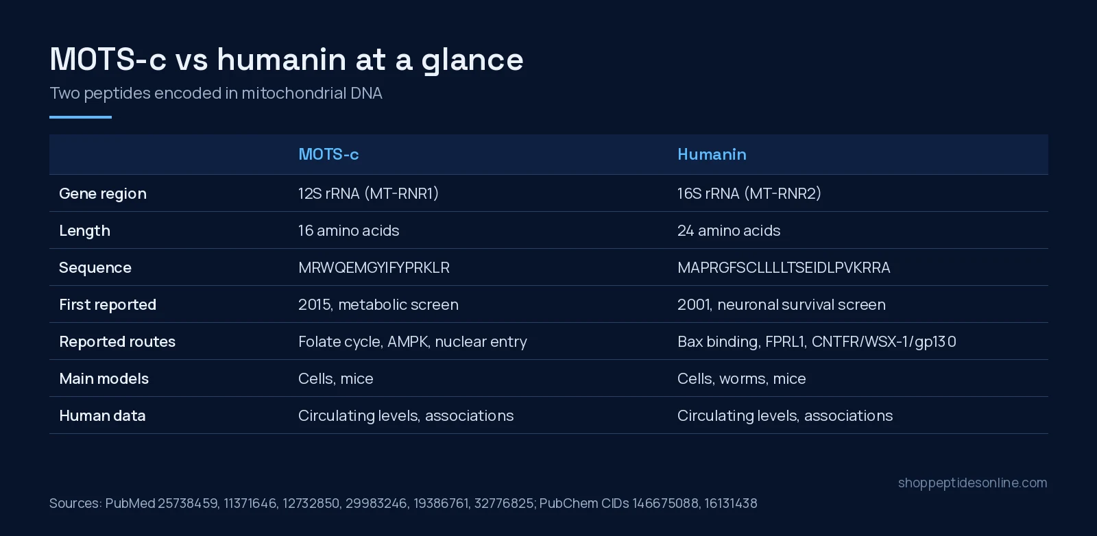
Human mitochondria carry a small circular genome. Most of it codes for respiratory-chain proteins, transfer RNAs and two ribosomal RNAs (12S and 16S). Short open reading frames tucked inside the two rRNA genes turned out to encode peptides, now grouped as mitochondrial-derived peptides (MDPs). A 2020 review counted eight: MOTS-c in the 12S rRNA gene (MT-RNR1), and humanin plus six small humanin-like peptides (SHLP1–6) in the 16S rRNA gene (MT-RNR2).[4],[5]
Humanin came first. In 2001, Hashimoto and colleagues ran a functional expression screen for genes that stopped neuronal cell death caused by familial Alzheimer’s disease genes and amyloid-beta. One hit encoded a short secreted polypeptide they named humanin.[2] Two years later, Guo and colleagues noted that the mitochondrial genome contains an identical open reading frame, and that the mitochondrial version also binds Bax.[3]
MOTS-c followed the lead. Lee and colleagues reasoned that if mtDNA held one signalling peptide, it might hold more. Their 2015 search turned up a 16-residue peptide inside the 12S rRNA gene, named MOTS-c (mitochondrial open reading frame of the 12S rRNA type-c).[1]
One complication applies to humanin and not, so far, to MOTS-c: the nuclear genome holds many mitochondria-like copies (NUMTs). Bodzioch and colleagues described 13 MT-RNR2-like nuclear loci predicted to encode humanin-like peptides, at least ten of them expressed in human tissues.[6] That makes it harder to say which gene a measured “humanin” signal came from.
Structure and identity
| MOTS-c | Humanin | |
|---|---|---|
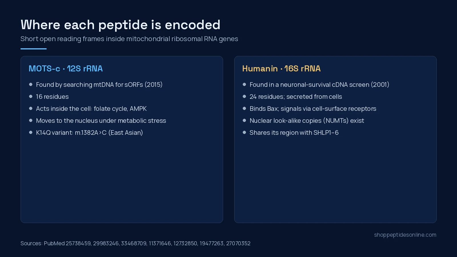
| Sequence | MRWQEMGYIFYPRKLR (16 aa)[1] | MAPRGFSCLLLLTSEIDLPVKRRA (24 aa)[3],[10] |
| Formula / mass (PubChem) | C101H152N28O22S2, about 2,175 Da (CID 146675088) | C119H204N34O32S2, about 2,687 Da (CID 16131438) |
| CAS number | 1627580-64-6 | 330936-69-1 |
| mtDNA gene | 12S rRNA (MT-RNR1)[4] | 16S rRNA (MT-RNR2)[4],[6] |
| Known human sequence variant | m.1382A>C, giving K14Q, found mainly in East Asian populations[15] | rs2854128 in the coding region, linked to lower circulating humanin[14] |
| Sibling peptides | None described in the 12S gene | SHLP1–6 in the same 16S region[5] |
How each is reported to act
MOTS-c: mostly intracellular metabolic signalling
In the 2015 discovery paper, MOTS-c inhibited the folate cycle and the purine synthesis tied to it, which led to AMPK activation in cells. Skeletal muscle appeared to be its main target tissue in mice.[1] A 2018 follow-up in cell models found that under glucose restriction and other metabolic stress, MOTS-c moved into the nucleus in an AMPK-dependent way and regulated nuclear genes, including antioxidant-response genes, interacting with the transcription factor NRF2.[7] These papers describe intracellular routes; they do not name a cell-surface receptor for MOTS-c.
Humanin: cell survival and surface receptors
Humanin’s best-known intracellular partner is Bax, a pro-apoptotic protein. In cell and cell-free experiments, humanin bound Bax, blocked its move from the cytosol to mitochondria and suppressed cytochrome c release; lowering humanin with siRNA had the opposite effect.[3] Humanin also binds IGFBP-3 with high affinity, through the binding protein’s heparin-binding domain.[10]
Outside the cell, two receptor systems have been reported. In 2004, synthetic humanin was shown to act on the G protein-coupled formyl peptide receptor-like 1 (FPRL1) on mononuclear phagocytes.[8] In 2009, neuroprotection by humanin in neuronal cells required gp130, and humanin drove the assembly of a complex of CNTF receptor alpha, WSX-1 and gp130.[9] Which receptor matters in which tissue is not settled.
What the animal and cell studies report
Most of what is known about both peptides comes from cultured cells and rodents. A summary, with the model always attached:
- MOTS-c, mice. In the discovery paper, MOTS-c treatment in mice prevented age-dependent and high-fat-diet-induced insulin resistance in those models.[1] A 2021 study reported better physical performance in young, middle-aged and old mice given MOTS-c, and changes in nuclear genes tied to metabolism and proteostasis.[11]
- Humanin, rodents. In rodent clamp studies, humanin infused into the brain ventricles improved overall insulin sensitivity, an effect tied to hypothalamic STAT-3 signalling.[12] In aged mice, humanin administration improved performance on cognitive tests.[14]
- Humanin, worms and transgenic mice. Overexpressing humanin in C. elegans extended lifespan in a daf-16/FOXO-dependent way, and humanin transgenic mice showed greater protection against toxic insults.[13]
- Humanin’s neighbours. Of the six SHLPs, SHLP2 and SHLP3 shared humanin’s protective effects on cell viability in vitro.[5]
None of this tells you what either peptide does in people. Rodent and worm results often fail to carry over, and the doses, routes and analogues used in these papers (for example the potent humanin analogue HNG[13]) are specific to each study.
What human data exist (observational only)
For both peptides, the human evidence is measurement and genetics: blood levels, muscle levels and gene variants. That evidence is associational, not proof of cause.
- Exercise. Exercise increased MOTS-c in human skeletal muscle and in circulation.[11]
- Age. Circulating humanin was lower in older humans and mice.[12] Children of centenarians had much higher circulating humanin than age-matched controls, and levels were lower in Alzheimer’s disease and MELAS.[13]
- Genetics. The MOTS-c K14Q variant was associated with type 2 diabetes prevalence in men, but not women, across three cohorts totalling 27,527 people.[15] A humanin-region variant (rs2854128) tracked with lower circulating humanin and with faster cognitive ageing in an independent cohort.[14]
- Vascular function. Among 40 patients having coronary testing, those with coronary endothelial dysfunction had lower plasma humanin.[17]
Null and contradictory findings
The record is not one-directional, and that matters when reading either literature.
- Plasma MOTS-c did not differ between lean and obese adults (10 per group). Its correlation with insulin-sensitivity indexes held only in the lean group.[16]
- In primary human fibroblasts driven into senescence, both humanin and MOTS-c were elevated, and adding either peptide modestly increased mitochondrial respiration and selected senescence-associated secretory factors, partly through JAK signalling.[18] That is the opposite of a simple protective story.
- In a 2026 study of adults with cerebral palsy and typically developing controls, circulating humanin and MOTS-c showed no or only modest changes after an acute endurance session.[19]
- The nuclear humanin-like copies mean that an antibody-based “humanin” measurement may not be reading the mitochondrial peptide alone.[6] The 2026 study, for example, measured MDPs with an in-house ELISA.[19]
Reviews in this field often end on optimism about MDPs as targets.[20] Treat that as a hypothesis the evidence above has not yet tested in people.
Which one fits a research question?
The two peptides answer different kinds of questions in the lab:
- Work on mitochondrial-to-nuclear signalling, AMPK and folate/purine metabolism in muscle cells is where most MOTS-c papers sit.[1],[7]
- Work on apoptosis, Bax, receptor signalling (FPRL1, gp130 complexes) and neuronal survival models is where most humanin papers sit.[3],[8],[9]
- Studies comparing several MDPs at once usually measure both, plus SHLPs, in the same samples.[18],[19]
Shop Peptides Online stocks MOTS-c for laboratory research, not humanin. The MOTS-c research hub lists the formats we carry, for example MOTS-c 10 mg. Third-party reports we have posted are on our COA page, listed by batch number. If you are comparing MOTS-c with a coenzyme rather than another peptide, see MOTS-c vs NAD+. For ordering and shipping, here is how to buy peptides online in Canada.
What the evidence does not show
- It does not show that either peptide, given to people, changes any health outcome. No interventional human data are cited here.
- It does not show a defined cell-surface receptor for MOTS-c.
- It does not settle which humanin receptor is relevant in which tissue, or how much measured humanin comes from nuclear look-alike genes.
- It does not show that lower blood levels cause any of the conditions they are associated with.
Frequently asked questions
What is the main difference between MOTS-c and humanin?
Both are short peptides encoded in mitochondrial DNA, but in different genes. MOTS-c is a 16-residue peptide from the 12S rRNA gene and is mainly described acting inside cells through the folate cycle, AMPK and the nucleus. Humanin is a 24-residue peptide from the 16S rRNA gene, first found as a neuronal survival factor, and is described binding Bax and signalling through cell-surface receptors.
Were MOTS-c and humanin discovered the same way?
No. Humanin was found in 2001 through a functional cDNA screen for factors that blocked neuronal cell death. MOTS-c was found in 2015 when researchers searched mitochondrial DNA for further short open reading frames after humanin.
Has MOTS-c or humanin been tested in human interventional trials?
The human data cited here are measurements of circulating levels and genetic associations, not trials of either peptide given to people. Neither peptide is an approved drug, and this article makes no health claims about either.
Do MOTS-c and humanin always act in the same direction?
Not always. In senescent human fibroblasts, both peptides modestly raised mitochondrial respiration and some senescence-associated secretory factors, which is not the protective picture reported in other models. Results depend on the cell type, model and condition studied.
Does Shop Peptides Online sell humanin?
No. Shop Peptides Online lists MOTS-c for laboratory research, not humanin. Humanin is covered here for comparison only.
References
- Lee C, Zeng J, Drew BG et al. (2015). The mitochondrial-derived peptide MOTS-c promotes metabolic homeostasis and reduces obesity and insulin resistance. Cell Metab. PMID: 25738459. doi: 10.1016/j.cmet.2015.02.009.
- Hashimoto Y, Niikura T, Tajima H et al. (2001). A rescue factor abolishing neuronal cell death by a wide spectrum of familial Alzheimer’s disease genes and Abeta. Proc Natl Acad Sci U S A. PMID: 11371646. doi: 10.1073/pnas.101133498.
- Guo B, Zhai D, Cabezas E et al. (2003). Humanin peptide suppresses apoptosis by interfering with Bax activation. Nature. PMID: 12732850. doi: 10.1038/nature01627.
- Merry TL, Chan A, Woodhead JST et al. (2020). Mitochondrial-derived peptides in energy metabolism. Am J Physiol Endocrinol Metab. PMID: 32776825. doi: 10.1152/ajpendo.00249.2020.
- Cobb LJ, Lee C, Xiao J et al. (2016). Naturally occurring mitochondrial-derived peptides are age-dependent regulators of apoptosis, insulin sensitivity, and inflammatory markers. Aging (Albany NY). PMID: 27070352. doi: 10.18632/aging.100943.
- Bodzioch M, Lapicka-Bodzioch K, Zapala B et al. (2009). Evidence for potential functionality of nuclearly-encoded humanin isoforms. Genomics. PMID: 19477263. doi: 10.1016/j.ygeno.2009.05.006.
- Kim KH, Son JM, Benayoun BA et al. (2018). The Mitochondrial-Encoded Peptide MOTS-c Translocates to the Nucleus to Regulate Nuclear Gene Expression in Response to Metabolic Stress. Cell Metab. PMID: 29983246. doi: 10.1016/j.cmet.2018.06.008.
- Ying G, Iribarren P, Zhou Y et al. (2004). Humanin, a newly identified neuroprotective factor, uses the G protein-coupled formylpeptide receptor-like-1 as a functional receptor. J Immunol. PMID: 15153530. doi: 10.4049/jimmunol.172.11.7078.
- Hashimoto Y, Kurita M, Aiso S et al. (2009). Humanin inhibits neuronal cell death by interacting with a cytokine receptor complex or complexes involving CNTF receptor alpha/WSX-1/gp130. Mol Biol Cell. PMID: 19386761. doi: 10.1091/mbc.e09-02-0168.
- Ikonen M, Liu B, Hashimoto Y et al. (2003). Interaction between the Alzheimer’s survival peptide humanin and insulin-like growth factor-binding protein 3 regulates cell survival and apoptosis. Proc Natl Acad Sci U S A. PMID: 14561895. doi: 10.1073/pnas.2135111100.
- Reynolds JC, Lai RW, Woodhead JST et al. (2021). MOTS-c is an exercise-induced mitochondrial-encoded regulator of age-dependent physical decline and muscle homeostasis. Nat Commun. PMID: 33473109. doi: 10.1038/s41467-020-20790-0.
- Muzumdar RH, Huffman DM, Atzmon G et al. (2009). Humanin: a novel central regulator of peripheral insulin action. PLoS One. PMID: 19623253. doi: 10.1371/journal.pone.0006334.
- Yen K, Mehta HH, Kim SJ et al. (2020). The mitochondrial derived peptide humanin is a regulator of lifespan and healthspan. Aging (Albany NY). PMID: 32575074. doi: 10.18632/aging.103534.
- Yen K, Wan J, Mehta HH et al. (2018). Humanin Prevents Age-Related Cognitive Decline in Mice and is Associated with Improved Cognitive Age in Humans. Sci Rep. PMID: 30242290. doi: 10.1038/s41598-018-32616-7.
- Zempo H, Kim SJ, Fuku N et al. (2021). A pro-diabetogenic mtDNA polymorphism in the mitochondrial-derived peptide, MOTS-c. Aging (Albany NY). PMID: 33468709. doi: 10.18632/aging.202529.
- Cataldo LR, Fernández-Verdejo R, Santos JL et al. (2018). Plasma MOTS-c levels are associated with insulin sensitivity in lean but not in obese individuals. J Investig Med. PMID: 29593067. doi: 10.1136/jim-2017-000681.
- Widmer RJ, Flammer AJ, Herrmann J et al. (2013). Circulating humanin levels are associated with preserved coronary endothelial function. Am J Physiol Heart Circ Physiol. PMID: 23220334. doi: 10.1152/ajpheart.00765.2012.
- Kim SJ, Mehta HH, Wan J et al. (2018). Mitochondrial peptides modulate mitochondrial function during cellular senescence. Aging (Albany NY). PMID: 29886458. doi: 10.18632/aging.101463.
- Horwath O, Corell L, Wan J et al. (2026). Circulating mitochondrial-derived microproteins at rest and in response to an acute bout of endurance exercise in individuals with cerebral palsy. Exp Physiol. PMID: 42349896. doi: 10.1113/EP093298.
- Kim SJ, Miller B, Kumagai H et al. (2021). Mitochondrial-derived peptides in aging and age-related diseases. Geroscience. PMID: 32910336. doi: 10.1007/s11357-020-00262-5.